Common Eye
Conditions in Adults
Common eye conditions
in adults
Pterygium
What is a pterygium?
A pterygium is an overgrowth of tissue from the white of the eye onto the clear cornea. It occurs due to a number of factors, with the most important being sunlight exposure during the early years of life. It is a common condition among young adults, especially in Queensland. A pterygium may remain stationary, or may continue to grow and extend further across the front of the eye. Such growth can cause redness, inflammation, discomfort and even disturbance to the quality of vision. In extreme circumstances, a pterygium may grow to cross the line of vision.
What is the treatment for a pterygium?
First and foremost, it is important to correctly diagnose pterygium. Changes similar to those seen in pterygium are possible in other conditions, and examination by an eye care professional is required to make the differentiation.
If a pterygium causes chronic redness, irritation, change to the vision, or if it changes suspiciously, treatment may be recommended. It may also be treated if its appearance is of cosmetic concern. Treatment approaches will depend upon individual factors, but may include:
- Avoidance of irritants
- Lubricating eye drops
- Sparing use of eye drops to treat redness or to provide temporary relief of symptoms
- Surgical removal
What is involved in pterygium surgery?
Your ophthalmologist will be able to provide you with advice regarding whether surgical treatment is recommended for your pterygium. The major complication of pterygium surgery is recurrence of the pterygium, however using modern meticulous techniques this is rare. Such an approach requires meticulous removal of the tissue responsible for causing the pterygium, followed by filling the resulting defect on the surface of the eye with graft of conjunctiva obtained from another part of the eye surface. Using this technique also provides the best possible cosmetic result.
The procedure is performed as day surgery under light sedation and local anaesthetic, and you are able to go home to recover overnight before being checked the next day by your ophthalmologist. The eye will be red and gritty for a few days after the surgery, and it is likely that it will take a few weeks to feel completely recovered. Eye drops are used during this time to help protect against infection, to settle inflammation and to aid healing.
Your ophthalmologist will monitor your recovery carefully, with 2-3 routine visits occurring within the months after surgery.
What can be done to prevent pterygia?
Exposure to sunlight during the early years of life, and ongoing exposure to sunlight throughout life, is an important factor in the development of pterygia. Therefore, sun protection in the form of wrap-around sunglasses, a broad-brimmed hat and avoiding sunlight during peak UV times from an early age are vital.
Cataract
Cataract is a cloudiness or loss of transparency of the lens in the eye. In most cases, cataracts cause blurring of vision, glare and decreased contrast under low lighting conditions and at night.
Some causes of cataract include:
- Congenital causes in infants include hereditary types, or secondary to infections or other medical conditions
- Metabolic conditions
- Age-related changes
- Diabetes
- Exposure to steroid medications
- Previous inflammatory conditions of the eye
- Previous injuries or operations on the eye
When a cataract starts causing problems with clear and comfortable vision, it may be appropriate to consider surgical removal and replacement with an artificial (intraocular) lens. At your appointment, your ophthalmologist will discuss your visual needs for work, hobbies and other tasks in order to help decide which intraocular artificial lens will suit you best. A series of measurements of your eye will be taken to facilitate this decision.
During cataract surgery, the cloudy crystalline lens is removed using microscopic ultrasound techniques, and replaced with a foldable artificial lens inserted through a very small wound in the eye. Usually no stitches are required.
A vast majority of cataract surgery is routine and successful. Complications are rare occurrences, and your ophthalmologist will discuss the risk of these with you at the time of your consultation, as each person’s risk is different.
Cataract surgery is a day procedure. Post-operatively, you will have a pad and shield over the eye for at least a few hours or overnight. You will be given eye drops to use in the eye for approximately four weeks, as prescribed. You will usually have a check-up appointment the day after your operation, and another within a few weeks. You will not be able to undertake any heavy lifting, driving or swimming until cleared by your ophthalmologist.
Strabismus
What is adult strabismus?
Strabismus (“turned eye”, “misaligned eyes”, “squint”)
Strabismus is a condition in which the eyeballs are not aligned properly and point in different directions. Nearly four in every 100 adults have this condition.
Adult strabismus results in visual, physical, and psychosocial disabilities. Successful strabismus surgery can relieve double vision and visual confusion, restore or establish depth perception, expand the visual field, eliminate an abnormal head posture, and improve psychosocial function and employability. Adults with strabismus should consult their ophthalmologist about the relative risks and benefits of surgery.
What causes strabismus in adults?
Most adults with strabismus have had the condition since earlier age in their life. However, strabismus can also begin in adulthood due to medical problems, such as:
- Eye muscle sagging with age
- Eye muscle nerve damage
- Thyroid disease (Graves’ disease)
- Myasthenia gravis
- Brain tumours
- Head trauma
- Stroke
- After cataract or retinal surgery
What are the symptoms of strabismus?
Adults with strabismus may experience:
- Eye fatigue
- Double vision
- Abnormal head turn
- A pulling sensation around the eyes
- Reading difficulty
- Loss of depth perception
These symptoms may have a negative impact on employment and social opportunities.
How is strabismus in adults treated?
Strabismus in adults can be treated using several methods, including:
- Eye patch (occlusion of one eye)
- Glasses containing prisms
- Eye muscle surgery
Eye Patch: This is simple and often resolve the double images instantly while investigations are undertaken, or recovery is anticipated.
Prism eye glasses: Eye glasses with prisms can correct mild double vision associated with strabismus in adults. A prism is a clear, wedge-shaped lens that bends, or refracts, light rays. When worn by an adult with strabismus who has mild double vision, the prism eye glasses realign images together so that the eyes see only one image. The prisms can be worn on the outside of the eye glass frames or can be manufactured directly into the lens itself. Prism eye glasses usually cannot correct more severe cases of double vision where images are far apart or double vision caused by weak or tight muscles.
Eye muscle surgery: Eye muscle surgery is the most common treatment for strabismus. Typically, strabismus occurs when the muscles surrounding the eyes are either too stiff or too weak. Our doctors can surgically loosen, tighten or reposition selected eye muscles so that the eyes can be rebalanced to work together.
Surgery for strabismus
Strabismus surgery can:
- Improve eye alignment
- Reduce or eliminate double vision
- Improve or restore the use of both eyes together (binocular visual function)
- Reduce eye fatigue
- Expand peripheral (side) vision
- Improve social and professional opportunities
Strabismus surgery is usually performed as a day procedure, using general or local anaesthesia. Patients may experience some pain or discomfort after surgery, but it is usually not severe and can be treated with over-the-counter pain medication such as paracetamol. Stronger medications for pain are sometimes needed and will be prescribed by your ophthalmologist or anaesthetist. You can often return to your normal activities within one to two weeks following surgery.
New strabismus procedures for complex strabismus
Surgery using adjustable sutures or eye muscle transposition is required in some cases with nerve damage, or previous unsuccessful eye muscle surgeries.
Valley Eye Specialists incorporates the Queensland Paediatric Ophthalmology and Strabismus Surgeons (QPOSS), a group of expert ophthalmologists who are trained in treating strabismus in adults, including complex strabismus.
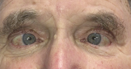
Pre-operative photograph – both eyes turned inward

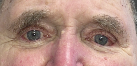
Post-operative photograph – both eyes are now straight

Glaucoma
Glaucoma is the name given to a group of eye conditions where vision is lost due to damage to the optic nerve, which functions like the battery pack of the eye. In most cases of glaucoma, there are no symptoms or warning signs in the early stages.
The primary issue in glaucoma is damage to the optic nerve due to a rise in intraocular pressure (IOP). This is the pressure of the fluid within the eye. The IOP at which progressive damage to the optic nerve occurs varies between different individuals. Thus, the best way to protect your sight from the effects of glaucoma is to have your eyes checked regularly. This is especially important as glaucoma cannot be self-detected, and people affected by glaucoma may not be aware of any vision loss.
Whilst there is no cure for glaucoma, and vision that has been lost cannot be restored, early detection and early initiation of treatment can arrest or significantly slow the progression of this condition.
Macular degeneration
The macula is a small region within the retina (the eye’s “camera film”) that is highly sensitive and produces detailed, colour images in our central field of vision.
Age-related macular degeneration (ARMD) occurs when the macula is damaged, affecting the ability to read, drive and engage in other activities requiring fine central vision. ARMD usually occurs in both eyes, but one eye may be worse than the other. It is a progressive condition, and early detection can improve the visual prognosis.
Common symptoms of macular degeneration include:
- A progressive decline in the ability to see objects clearly
- Distorted vision
- Dark spots obscuring central vision
- Diminished colour appreciation
When complications of ARMD cause substantial disturbances in vision, it is termed “late ARMD”. There are two types of late ARMD:
- Dry ARMD – this is usually slower to develop than the wet form (see below). There is no approved treatment for ARMD, but studies are underway investigating disease progression and potential future therapies.
- Wet ARMD – this is when new, abnormal blood vessels grow from the layers underneath the macula. These new blood vessels are fragile and tend to leak and bleed, resulting in severe distortion of vision and eventually formation of scar tissue. Whilst there is no cure for wet ARMD, expert treatment can slow or even stop the progression of the disease, maintaining (or even improving) vision for many patients.
Valley Eye Specialists is equipped with the latest technology to enable early detection and correct diagnosis of macular degeneration. Urgent treatment for wet ARMD can be provided in our practice, and we strive to make appointments available for same day referrals if needed.
Diabetic eye conditions
Diabetes can damage the blood vessels at the back of the eye, resulting in a number of problems.
Diabetic maculopathy
Damaged blood vessels may leak blood and fluid into the retina, which causes swelling. When this process occurs at the macula, vision may become blurred or distorted.
Diabetic retinopathy
Further damage results in closure of blood vessels within the retina, starving it of the oxygen which is needed for it to function properly and provide clear vision. The eye attempts to compensate for this by growing new blood vessels. However, these new blood vessels are fragile, leaky and often grow in abnormal locations. They can bleed and create scar tissue, resulting in vision loss.
When should I have my eyes checked for changes due to diabetes?
The chance of developing changes in the the eye due to diabetes increases with time, and is more likely if blood sugar levels are poorly controlled or if other problems such as high blood pressure and high cholesterol are present.
Early diagnosis and careful follow-up are critical in preventing vision loss due to diabetic eye conditions. However, affected people are often unaware of the early changes that occur due to diabetes. Diabetic changes are not painful and the external appearance of the eye is does not change. Macular changes in one eye may not be noticed, as the unaffected eye can readily compensate for the reduction in vision. Diabetic retinopathy is usually asymptomatic in the early stages, resulting in changes to vision only once complications have occurred. This means that routine eye checks are vital. For those with type 1 diabetes, an initial eye examination should occur within five years of diagnosis. For those with type 2 diabetes, this should occur around the time of diagnosis. Even if no changes are evident, reviews at least every two years are required.
How are diabetic eye conditions diagnosed?
A thorough check up involves dilating the pupils with eye drops and a careful examination of the inside of both eyes. A macular scan known as an OCT (ocular coherence tomography) will be performed. This is a simple and painless test where the macula is scanned by a laser beam to produce a detailed map of this area which can then be closely reviewed to detect early changes or monitor progression of changes. High-resolution colour photographs of the retina will also be taken, and these will help to determine the severity of any changes present as well as allow monitoring of the eye’s health over time.
In some circumstances, a fluorescein angiogram may be recommended. This provides additional information about the macula and retina, and can better show the extent and severity of damage. It can also help determine when laser treatment is needed and can help to show where such laser treatment should be directed. This test is made possible through the injection of a small amount of dye called fluorescein into an arm vein. Retinal photographs are then taken in rapid succession, using a special filter that helps to highlight the dye as it circulates throughout the blood vessels at the back of the eye.
After your examination and review of the additional scans and imaging performed, your ophthalmologist will be able to advise you whether you have any diabetic eye changes, and will discuss the need for further monitoring and/or treatment.
What treatment may be required?
Currently available treatment options include injection of therapeutic medications into the vitreous cavity (back) of the eye, laser treatment and surgery. Your ophthalmologist will consider factors such as your level of vision, age, diabetic severity, other medical issues, and degree of diabetic eye changes when determining whether any treatment is required. In many cases, even if changes are detected, treatment is not necessary. However, regular and careful examinations must be continued.
Blepharitis
Blepharitis is a chronic inflammatory condition involving the eyelid margins. If can affect both children and adults. Mostly, it responds to simple treatment and is not a vision-threatening problem.
Blepharitis arises from blocked glands (Meibomian glands) and an alteration in the substance that these glands normally make and secrete onto the margin of the eyelid. It is thought that, in some people, blepharitis is caused by a sensitivity to the bacteria which normally live on the skin.
Common symptoms of blepharitis include:
- Red eyelids
- Crusting of the eyelashes
- Red, dry or irritated eyes
The most important treatment for blepharitis is good “eyelid hygiene”, and this should be performed regularly even when eyes seem comfortable and uninflamed. Other treatments may be required if this is not effective on its own, and your ophthalmologist will be able to discuss which treatments will be most likely to help in your particular case.
When untreated or severe, blepharitis may lead to complications such as:
- Chalazion
- Chronic conjunctivitis
- Tear film abnormalities like dry eyes
- Marginal keratitis
- Loss of eyelashes
- Scarring
These conditions are best treated by your ophthalmologist, but most of the time prevention is possible through good eyelid hygiene.
Chalazion
A chalazion, or Meibomian cyst, is a common eye condition in both children and adults alike. It usually presents as a lump on the eyelid which waxes and wanes with time. These lumps can be single or multiple and may affect one eye or both simultaneously or subsequently. People with conditions such as rosacea, seborrheic dermatitis or blepharitis are more prone to multiple and recurrent chalazia.
Chalazion arises from a blocked gland (Meibomian gland) that is present in the normal eyelid. These blocked glands then become chronically inflamed, and enlarge to form a painless lump at the eyelid margin. These lumps are non-infective and non-contagious, but on occasion secondary infection is possible.
Mostly chalazia respond to simple eyelid hygiene practices involving regular daily warm compresses and massaging the lid margins. If complicated by infection, antibiotics are usually also required. Rarely, chalazia will need to be treated with a simple surgical drainage procedure if they are very large, chronic, infected or do not respond to the simple treatments outlined above.
Eyelid lesions
Living in the beautiful Sunshine State, we see many eyelid lesions, both benign (non-cancerous) and malignant (cancerous). The eyelids are highly susceptible to sun damage and thus, skin cancer. Any changes in the appearance of the eyelid skin (swelling, thickening, ulceration) or the presence of a new lesion requires prompt evaluation and, if necessary, early intervention.
Eyelid malposition
Eyelid malposition (ectropion and entropion) occurs when the eyelid is no longer positioned against the eyeball as it should be. The malposition is called an “ectropion” if the eyelid turns outwards, and an “entropion” if it turns inwards. Both conditions are usually (but not always) associated with ageing, and the effects of sun exposure, and may lead to watering, redness and discomfort. There are different ways to treat eyelid malposition, depending on the underlying cause, including non-surgical and surgical measures.
Dermatochalasis
Excess upper eyelid skin (dermatochalasis) may warrant treatment with surgery if the skin is affecting vision or making the eyelids heavy and difficult to keep open. Careful consideration is given to ensure there is no underlying brow or eyelid malposition, so that the appropriate procedure (blepharoplasty) is performed.
Ocular side effects of medications
A number of medications, prescribed for various conditions, have the potential to cause unwanted effects on the eye and vision. Most often, with appropriate screening and monitoring, such side effects can be detected early before they cause permanent changes. Examples of common medications that may require monitoring by an optometrist or an ophthalmologist include:
- Plaquenil (hydroxychloroquine)
- Sabril (vigabatrin)
- Gilenya (fingolimod)
- Some specialised antibiotics (ethambutol and isoniazid)
The types of problems such medications may cause vary, and the risk to an individual of developing visual problems depends on a multitude of factors. Our ophthalmologists know how to appropriately screen for and detect early signs of medication-related eye changes in both adults and children, and can advise regarding ongoing monitoring requirements or liaise with your prescribing doctor to ensure ongoing risks are minimised.


